Study involving MAPI on the impact of AI bounding box display on radiologists’ interpretation published in European Journal of Radiology Open

Medical AI Promotion Institute Co., Ltd. (Head Office: Chuo-ku, Tokyo; Chief Executive Officer: Yuki Shimahara; Representative Director: Sho Kawabe; hereinafter “MAPI”) announces that research in which MAPI participated has been published in the international academic journal European Journal of Radiology Open. This study quantitatively evaluated, using eye-tracking technology, how AI-assisted lesion candidate highlighting (bounding box display (*1)) influences radiologists’ eye movements and interpretation behavior when reading chest X-ray images.
Background
In recent years, AI-assisted diagnostic systems (CAD) that present lesion candidates using bounding boxes and other visual indicators have been widely adopted in clinical radiology, including chest X-ray interpretation. While these displays are useful as diagnostic support tools, there have been concerns that they may also influence the radiologists’ interpretation process itself. Therefore, this study investigated how CAD displays affect visual search behavior during image interpretation using eye-tracking technology.
Overview
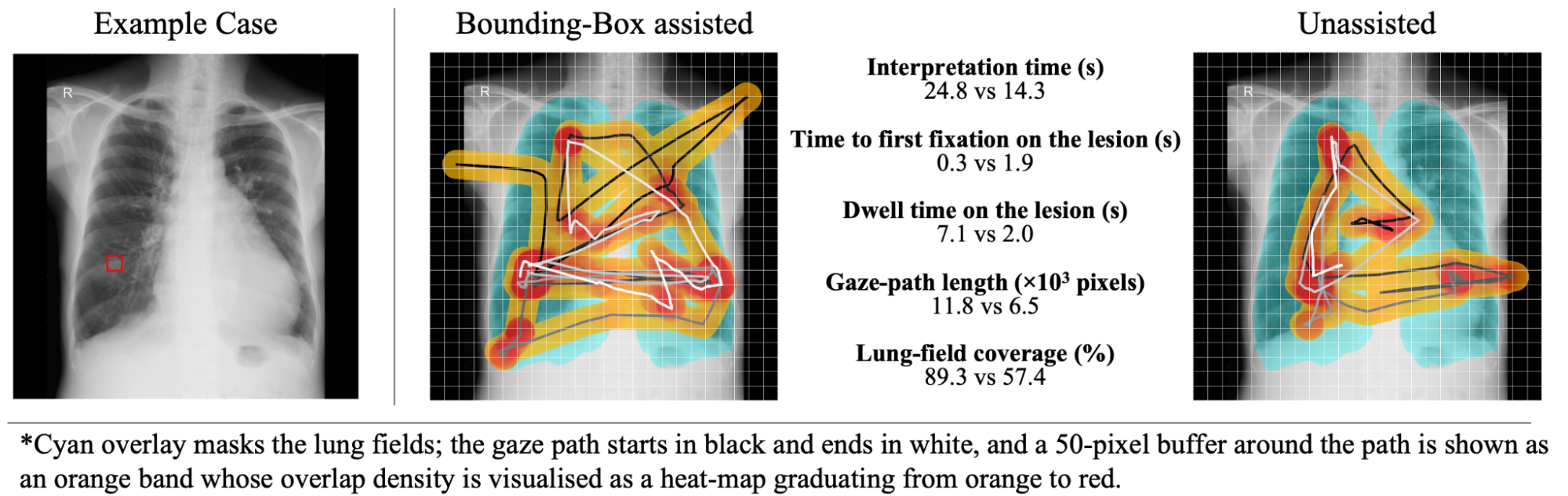
A total of 180 chest X-ray images (including cases with lung nodules or masses and normal cases) selected from a public dataset were interpreted twice by three radiologists, with and without lesion candidate bounding boxes. In the bounding box condition, a “Concurrent Reader” format was used, in which the bounding boxes were displayed immediately upon opening the image.
Eye movements during interpretation were recorded using the EyeTech VT3 Mini (*2). Metrics analyzed included total interpretation time, time to first fixation on the lesion, lesion fixation duration, total eye movement distance, and coverage rate of the lung field.
Main Results
When bounding boxes were present, the radiologists’ interpretation time increased on average, along with lesion fixation duration, eye movement distance, and lung field coverage. Meanwhile, the time to first fixation on the lesion was reduced, indicating that AI display influenced visual attention guidance.
These findings suggest that lesion candidate display affects the radiologists’ interpretation process.
Significance
This study demonstrated that, beyond conventional evaluation of diagnostic accuracy, eye-tracking enables visualization of how radiologists observe images and reach diagnostic decisions. It highlights the importance of evaluating not only whether AI is accurate, but also how it is used and how it influences human interpretation behavior. MAPI remains committed to advancing research on human–AI interaction to support the safe and appropriate clinical implementation of AI technologies.
Publication Information
Journal: European Journal of Radiology Open
Title:
Eye tracking as a tool to quantify the effects of CAD display on radiologists’ interpretation of chest radiographs
Year: 2026
Authors:
Daisuke Matsumoto, Tomohiro Kikuchi, Yusuke Takagi, Soichiro Kojima,
Ryoma Kobayashi, Daiju Ueda, Kohei Yamamoto, Sho Kawabe & Harushi Mori
DOI: 10.1016/j.ejro.2026.100731
URL:
https://www.sciencedirect.com/science/article/pii/S2352047726000080
(*1) A display method that highlights specific regions (such as lesions) with rectangular bounding boxes.
(*2) A stationary, non-contact eye-tracking device that measures gaze position and movement.
Company Profile
Company Name: Medical AI Promotion Institute Co., Ltd.
CEO: Yuki Shimahara, Sho Kawabe
Address: 4F Life Science Building 9, 12-9 Nihonbashi-Odenmacho, Chuo-ku, Tokyo, Japan
Established: November 8, 2023
Business: Development and operation of next-generation medical database business
Website:
https://mapi-jp.org
Facebook:
https://www.facebook.com/iryoaisuishinkiko
X:
https://twitter.com/iryoai_mapi
Contact
Public Relations
Medical AI Promotion Institute
Email: pr@mapi-jp.org